
Description
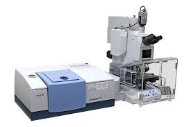
A gel documentation system combines a high-resolution digital imaging device with a darkroom enclosure, transilluminator, and software interface. Its main function is to photograph or scan electrophoresis gels that contain DNA, RNA, or proteins stained with dyes like ethidium bromide, SYBR Safe, GelRed, Coomassie Brilliant Blue, or silver stain. These dyes bind to the biomolecules and emit light (usually in the UV or visible range), allowing the bands to be detected and recorded.
The typical components of a gel doc system include:
1. Darkroom Enclosure:
A light-tight cabinet that protects the gel from ambient light and protects the user from UV exposure. It creates optimal imaging conditions.
2. Transilluminator:
A light source, often ultraviolet (UV), blue light, or white light, used to illuminate the gel from below or above, depending on the system and dye type.
3. Camera System:
A high-resolution digital camera (often CCD or CMOS) mounted above the gel records the fluorescence or coloration of bands with great sensitivity and clarity.
4. Imaging and Analysis Software:
Software is used to control the camera, adjust exposure, and process images. It also allows quantitative analysis, such as calculating molecular weights, estimating DNA/protein concentration, and comparing band intensity.
5. Filters and Lenses:
Optical filters are used to block unwanted wavelengths of light and enhance the visibility of fluorescent signals. Lenses focus the image onto the camera sensor.
—
Applications:
DNA/RNA analysis: Visualizing PCR products, restriction digests, and nucleic acid quantification.
Protein analysis: Imaging SDS-PAGE gels stained with Coomassie, silver stain, or fluorescent dyes.
Documentation: Archiving gel images for publications, lab reports, or quality control.
Quantitative analysis: Measuring band intensity to estimate the concentration or relative abundance of nucleic acids or proteins.
Verification: Confirming successful amplification or digestion before downstream applications (e.g., cloning, sequencing).
—
Advantages:
High sensitivity for detecting low-abundance samples.
Digital image capture allows for easy storage, sharing, and publication.
Quantitative analysis tools aid in precise measurements.
Minimizes user exposure to harmful UV radiation (especially with blue light systems).
Versatile imaging for a variety of gel types and stains.
—
Limitations:
UV light can damage nucleic acids if exposure is prolonged.
Ethidium bromide, a common stain, is mutagenic and requires proper handling and disposal.
High-resolution systems can be expensive.
Fluorescence-based detection may require specific filters and careful calibration.
—
Recent Developments:
Modern gel doc systems have evolved with the integration of:
Touchscreen controls
Wi-Fi/USB data export
Automated exposure adjustment
Blue/green light transilluminators (safer alternatives to UV)
Portable models for field use or mobile labs
—
In summary, the gel doc system is a cornerstone of molecular biology labs, enabling scientists to visualize and document biomolecular separation results quickly, safely, and accurately. Its integration with digital technology enhances both research productivity and data reproducibility.

Reviews
There are no reviews yet.